Welcome to the LST-AI segmentation service in neuGRID!
How to run the segmentation on MRI scans:
You have to upload:
i. Zip file containing DICOMS
or
ii. nii.gz archive
Then provide the following information:
- Age of the patient
- Sex of the patient (Male or Female)
- Normative reference curves (Note: this will modify the normative curves in the PDF report. If you choose M: they will be from the male healthy population; F: from the female healthy population; BOTH: composed of both male and female healthy controls)
- Flair type (2D or 3D)
and click the submit button!
LST-AI – Deep Learning Ensemble for Accurate MS Lesion Segmentation – segments T2 hyperintense lesions in FLAIR images. LST-AI is an advanced deep learning-based extension of the original LST toolbox. LST-AI was trained using an ensemble network model with data from 491 Multiple Sclerosis patients with severe lesion patterns. It also labels lesions such as periventricular, infratentorial, juxtacortical, and subcortical according to the 2017 McDonald criteria. Further information on the algorithm can be found in the publication by Wiltgen, 2024.
The LST-AI pipeline implemented in neuGRID requires FLAIR and T13D images of the subject. Lesion volume values are corrected by dividing by the subject’s Total Intracranial Volume, calculated by SPM12, and multiplying by a constant.
Once the LST-AI algorithm execution is complete, the outputs are stored in a PDF report showing the number and volume of lesions, the segmentation of lesions in relevant brain scans, and the subject’s position in the Prediction Percentile plot for white matter lesion volume. Each coloured line in the normative plot represents the Prediction Percentile for white matter lesion volume of a non-pathological subject; each number in the legend indicates the percentage of non-pathological subjects with a white matter lesion volume below the respective coloured line. If the subject is located above the 95% coloured line, the subject should be considered pathological.
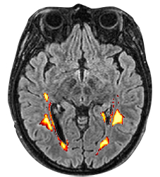
Probabilistic lesion volume map generated by LST-AI on a FLAIR image in axial view.